RUDN chemist created a new fluorescent probe for glutathione detection

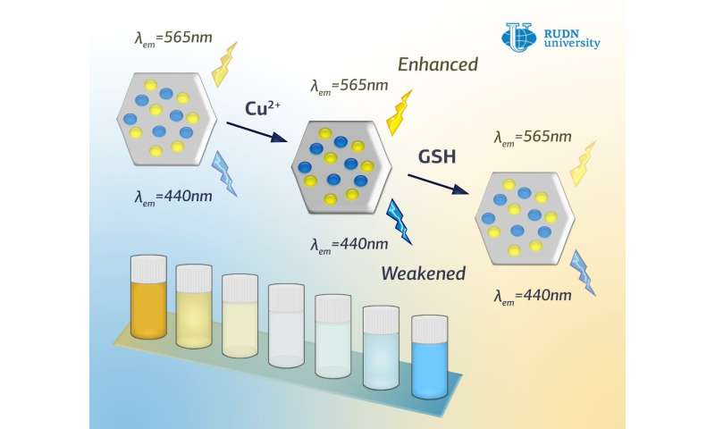
RUDN chemist in cooperation with scientists from Tabriz University (Iran) and Cordoba University (Spain) created metal-organic framework material with double emission that can be used as a fluorescent probe for the detection of glutathione, one of the most complex biological molecules. The method is based on the analysis of carbonic, not quantum dots. Due to the probe's high sensitivity towards glutathione, a molecule can be seen with naked eye, as the color changes from yellow to blue. The article is published in the journal Sensors and Actuators B: Chemical
Glutathione is a complex molecular compound, responsible for regulation of physiological processes in a cell. Ration between oxygenated and reduced molecules of glutathione shows the scale of detoxication in the body. Blood glutathione test helps diagnose susceptibility to serious diseases, such as Alzheimer's and Parkinson diseases. There are a number of methods to assess the level of glutathione, however, most of them are complicated, expensive, and not accurate enough. Moreover, there are structurally matching molecules, such as cysteine and homocysteine, which makes it difficult to detect glutathione.
Until now the whole range of measurement methods of concentration in fluorescent analysis has been based on quantum dots (QD) analysis—crystals of semiconductor, where quantum effects occur. Quantum dots can be used to construct multicolour fluorescent systems because of their continuous spectrums. Nevertheless, the high cost of QD and toxic elements such as cadmium and lead limit their usage, especially for biomedical research.
Carbon dots are a new type of nanomaterial, characterized by compatibility, adjustable photoluminescence, and low cytotoxocity. Carbon dots are the best alternative to traditional quantum dots in the area of biological system and biostructures visualization. However, most of the known carbon dots show radiation with higher wavelength. Section's colour tuning has been rarely tested, that is why very few fluorescent probes based on carbon dots are known.
To create a fluorescent probe for glutathione detection, Professor Rafael Luque, director of Scientific Centre of Molecule Design and Synthesis of Innovation Materials for Medicine of United Institute of Chemical Research at RUDN was the first to suggest using two forms of carbon dots at the same time: with yellow and blue emitting power, embedded in a zeolit-imidazole matrix cavity. This synthesis process allows to obtain material with advanced radiation in a visible range by fluorescent coefficient correlation modification at 565 and 440 nm sites.
"We were able to create a new double beam probe by encapsulating yellow and blue emitting carbon dots in the cavity of metal-organic framework. As a result of exposure of glutathione molecules with copper ions, that are at the structure of derived composite, intensive suppression of radiation from yellow carbon dots and recovery of radiation from blue dots occurs. Thus correlation modification of two radiations results in a distinctive change of fluorescent colour from yellow to blue. The high sensitivity of derived composite used as a probe has been achieved by high and selective accumulation of target analyte, in this case glutathione in its porous structure, which enables avoiding potential disturbances of coexistent materials. Apart from the fast visual identification of glutathione, the created probe is capable to identify molecules' content in the range 3–25 nm with a very low detection level 0,90 nm", the authors write.
Professor Luque and his colleagues conducted experiments for identifying glutathione in grape and cucumber specimens to test the precision and applicability of the probe on real biological objects. Glutathione was added to the fruit specimens and then tested by reduction fluorescence method, which is the method to measure the speed that a certain molecule (glutathione) interconnects with other similar molecules in the area of a cell. The obtained signal of fluorescence ranged from 94,2% up to 105,6%, which proves the ability of the fluorescent probe to selectively identify glutathione in the real specimens.
Among the advantages of the probe are not only the detection of the target molecule at low levels, but also that it is obtained by environmentally-friendly, cost-effective, and simple methods. The strategy for creating the probe may become a universal platform in the process of developing fluorescent sensors for identifying other molecules.
Article:
Dual-colored carbon dot encapsulated metal-organic framework for ratiometric detection of glutathione
Sensors & Actuators: B. Chemical
Volume 297, 2019, 126775
DOI: 10.1016/j.snb.2019.126775
https://www.sciencedirect.com/science/article/pii/S092540051930975X?via%3Dihub
Research area: materials chemistry, biotechnology
People's Friendship University of Russia (RUDN University)
Provided by RUDN University